For Appointment in Circle health Group Hospitals TEL: 08081010337, For Timings Click here.
SHOULDER PROBLEMS
Shoulder (subacromial) impingement
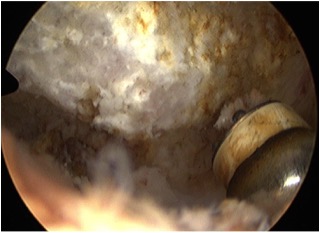

What is it? Shoulder joint is wrapped by a group of muscles called rotator cuff. These muscles form a tendon and are attached to the top of the arm bone (humeral head). The acromion , which is a part of shoulder blade bone, along with a ligament forms a roof over the humeral head and the rotator cuff tendon. As you lift your arm the tendon gets squashed between a spur arising from the acromion or from a thickened ligament. Overlying the rotator cuff tendon is the thin layer of tissue called the bursa, which normally helps in the gliding of the tendon during shoulder movement. This bursa gets inflamed and also causes pain.
Do I need more tests? You may require plain X—ray of the shoulder followed by Ultrasound scan or an MRI scan
What is the treatment? Analgesics and rest followed by physiotherapy is the usual initial treatment. You may benefit from a steroid injection into the shoulder. If this is not successful then you may need surgical treatment. Arthroscopic subacromial decompression is the surgical procedure.
What is arthroscopic subacromial decompression? This is a minimally invasive surgery performed arthroscopically under general anesthesia. The entire shoulder joint is inspected and all other abnormalities are addressed. Surgical instruments are introduced into the joint to smoothen the bone spurs on the acromion and/or collarbone
Rotator cuff tear
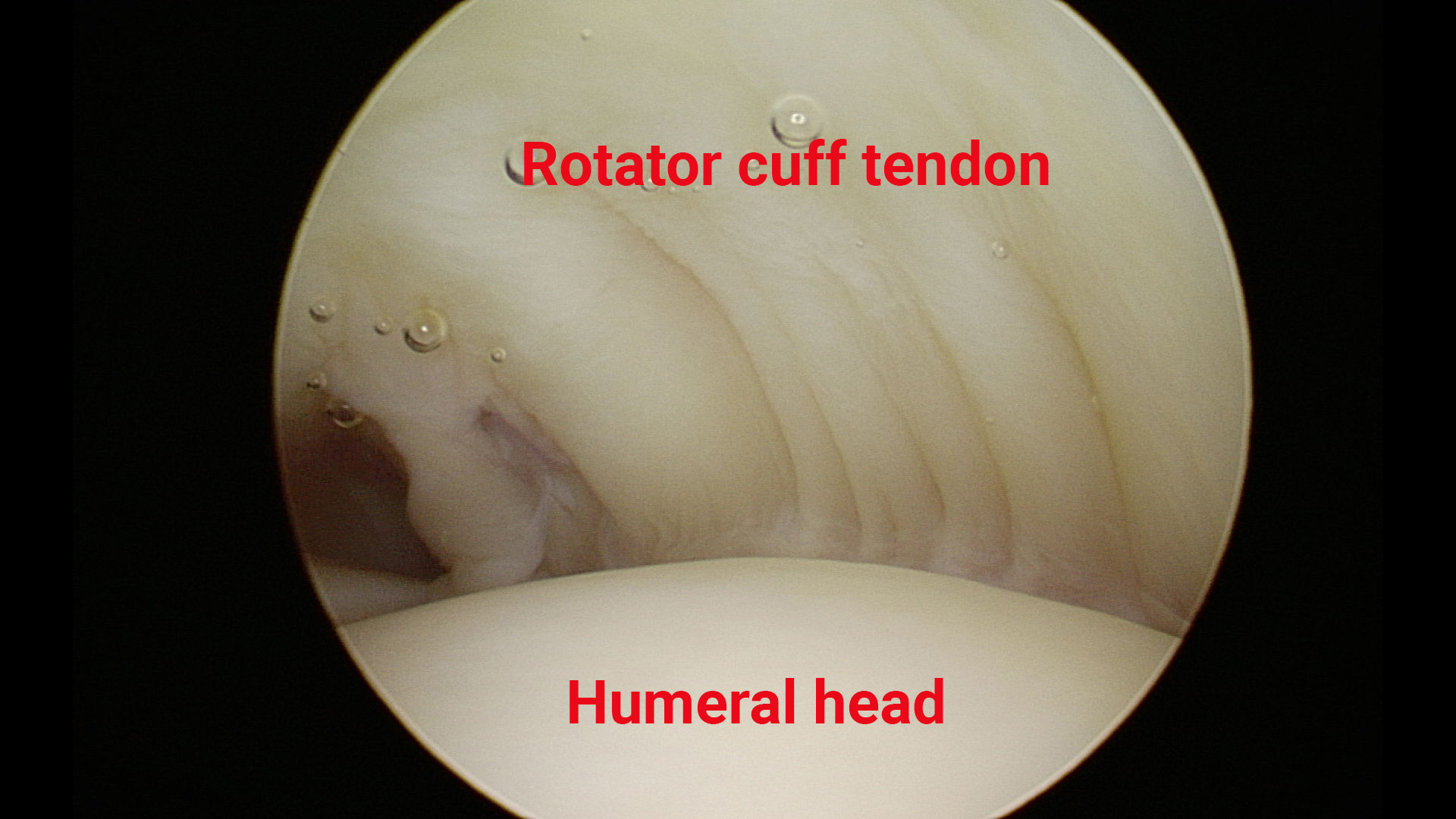
What is it? Shoulder joint is wrapped by a group of muscles called rotator cuff. These muscles are attached to the shoulder blade and form a tendon and are attached to the top of the arm bone (humeral head). These tendons can get torn either due to an injury (acute tear) or due to long-term wear and tear (chronic tear). Rotator cuff is important for the normal function of the shoulder joint and tendon tear can lead to pain and weakness of the arm.
Do I need more tests? You may require plain X—ray of the shoulder followed by Ultrasound scan or an MRI scan
What is the non surgical treatment? This can be treated symptomatically using painkillers, injection and physiotherapy. Full thickness rotator cuff tear does not heal naturally however it may become less symptomatic and shoulder function might compensate eventually with non-surgical treatment. There is a possibility that tear may become bigger over a period of time.
What is the surgical treatment? Rotator cuff tear may be amenable to repair using arthroscopic surgery. This is performed using small puncture wounds on the skin, which would allow insertion of a camera and instruments into the shoulder. The torn tendon is reattached to the bone using an anchor with sutures, which is passed through the torn tendon ends.
Calcific tendonits

What is it? This is a clinical condition characterized by deposition of calcium in the rotator cuff tendon and the reason for this is unknown. This causes severe pain over the shoulder and can leads to irritation of the tendon and inflammation of the overlying bursa.
Do I need more tests? Plain X—ray of the shoulder may show calcium deposits and some times further imaging is required in the form off Ultrasound scan or an MRI scan
What is the treatment? This can be treated by nonsteroidal anti-inflammatory medications to reduce the pain . A steroid injection may be useful to reduce the inflammation of the bursa followed by physiotherapy to strengthen the rotator cuff. If the non-surgical treatment is unsuccessful then a key hole surgery of the shoulder may be beneficial. Surgery is performed using an arthroscope and it involves a subacromial decompression and excision of the calcium deposits.
Click here for Patient Information Leaflet
Acromio-clavicular(AC) joint arthritis
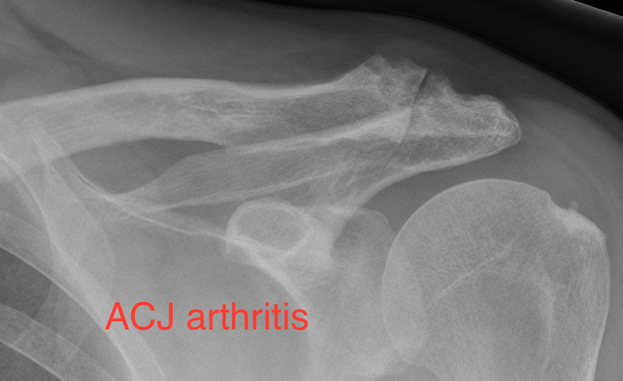
What is it? AC joint is formed by the outer end of the collarbone and top end of the shoulder blade (acromion). AC joint arthritis is usually caused by the constant wear and tear and there may be a history of injury in the past. This can lead to pain over the top of the shoulder causing difficulty in raising the arm high or across the body.
facharbeiten kaufenDo I need more tests? Plain X-ray would help in the diagnosis, further imaging is required if there is any other suspected pathology.
What is the treatment? Painkillers can help to treat this condition. A steroid injection into the joint may help to reduce the symptom. Surgical treatment involves excising the outer end of collarbone using a burr and this can be performed by a keyhole technique with the help of an arthroscope.
Biceps tendonits
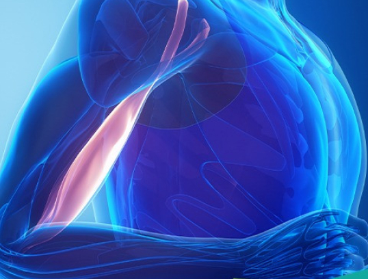
What is it? This is the inflammation of the long head of the biceps tendon, which passes through the shoulder joint before joining with the short head to form the biceps muscle. This can cause pain in the front of the shoulder with worsening of symptoms while lifting weight over the head especially in the front of the body. This is caused by wear and tear or an injury.
Do I need more tests? Ultrasound scan is helpful in diagnosing this condition. MRI scan can also be used.
What is the treatment? Rest, anti-inflammatory medicines and physiotherapy can treat this. A steroid injection may be given under ultrasound guidance to reduce inflammation and pain. If symptoms persist then surgical treatment may be undertaken. The surgery involves either a Tenotomy (releasing the tendon inside the shoulder joint) or tenodesis ( releasing the tendon inside the shoulder joint followed by reattaching the tendon to the top of the arm bone). Tenotomy is done by keyhole surgery. Tenodesis is either done by a keyhole surgery or a combination of keyhole and mini open procedure. Tenodesis is preferred for young active patients.
Frozen shoulder
What is it? This is a painful condition of the shoulder joint leading to stiffness due to inflammation and thickening of the capsule of the joint. This is often spontaneous in origin but can be due to injury, immobilisation or surgery of the shoulder joint. This is also seen more in patients with diabetes.
Do I need more tests? Plain X-Ray of the shoulder is performed to rule of arthritis of the shoulder joint.
What is the no surgical treatment? This may get better spontaneously but can take several years. The treatment options include painkillers, physiotherapy and shoulder joint injections with steroids.
What is the surgical treatment? If the above treatment fails then surgery is considered. This is called arthroscopic capsular release. This is a key hole procedure performed under general anaesthesia in which the capsule of the shoulder joint is release using instruments introduced into the shoulder joint through small puncture wounds.
Shoulder dislocation
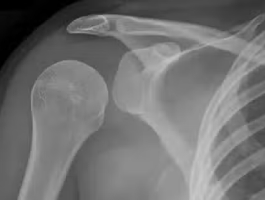
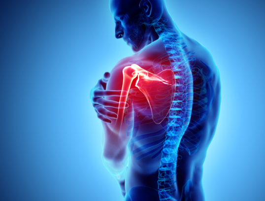
What is it? Shoulder is a very mobile ball and socket joint. The socket is very shallow compared to the size of the ball. There is a rim of tissue called labrum that increases the depth of the socket providing stability. The capsule, ligaments and muscle around the shoulder joint also provide stability. The ‘ball’ can come out of the socket commonly due to injury especially in young adults playing contact sports. The labrum can get torn off the socket during this process and this can lead to recurrent dislocation especially in young adults.
Do I need more tests? Plain x-ray of the shoulder joint is performed before and after reduction. In case of recurrent dislocation more detailed test in the form of MRI arthrogram (dye injected into the shoulder at the time of scan) is carried out to access the damage to the labrum.
What is the treatment? Once the shoulder joint is reduced, a short period of rest is recommended in a sling followed by physiotherapy. In young and active adults especially who play high level of sports, arthroscopic surgery to repair the torn labrum is recommended after the initial dislocation. Patients who are suffering from recurrent dislocation following the traumatic shoulder dislocations would also benefit from surgery. If the arthroscopic surgery to repair the labrum is not successful then it may be necessary to perform a bony procedure to prevent recurrent dislocation.
Click here for Patient Information Leaflet
SLAP tear
What is it? The socket of the shoulder joint has a rim of tissue at the margin called the labrum. SLAP tear is essentially the tear involving the superior (top) labrum, which has the attachment of longhead of the biceps tendon. SLAP stands for superior labrum anterior (front) to posterior (back). This is commonly seen after injury and this can leads to shoulder pain especially with overhead activities.
Do I need more tests? MRI arthrogram (dye injected into the shoulder at the time of scan) is carried out to look at damage to the labrum.
What is the treatment? Non-operative treatment with painkillers and rest can be tried initially but if it doesn’t settles then this may need shoulder arthroscopic surgery. The torn labrum is repaired with sutures and anchor placed at the top edge of the glenoid (socket) rim. In older patients long head of biceps tendon is detached and if necessary reattached to the top of the arm bone either using an anchor or an endo button.

Acromio-Clavicular (AC) Joint Dislocation

What is it? AC joint is formed by the outer end of the collarbone and top end of the shoulder blade (acromion). This joint can be injured due to fall on to the shoulder or on an outstretched hand. The ligaments surrounding this joint gets damaged and in severe cases the ligaments on the undersurface of the collar bone get ruptured leading on to separation of collarbone from the shoulder blade. AC joint pain and swelling is seen in acute injuries. In severe cases marked deformity (bony lump) is evident.
Do I need more tests? Plain X-Ray is enough to diagnosis and also to grade the severity of this injury
What is the treatment? Acute injury can be treated by immobilisation in a sling, painkillers and local icepack application. Once the pain improves, formal physiotherapy will be beneficial. Pain and loss of function can improve almost fully with nonoperative treatment however deformity will be permanent. In some patients pain and weakness persists and then surgical treatment may be considered. In severe cases causing complete disruption of ligament, surgical treatment may be carried out to provide stability to the joint and reducing the deformity. There are various surgical techniques described and my preferred technique is using a synthetic ligament to reconstruct the ligaments.
Fractures of the shoulder and clavicle
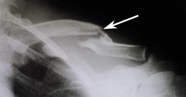
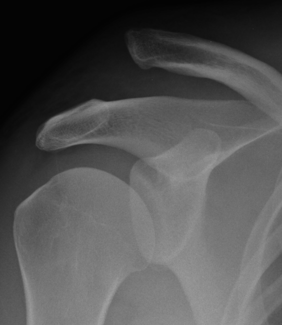
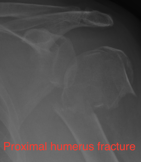
What are the common fractures affecting shoulder? Shoulder joint is a ball and socket joint. Fracture of the head of the humerus (ball) is one of the common fractures of the shoulder. These fractures tend to occur more commonly in older patients due to low velocity injuries. Fractures involving the socket and shoulder blade are rare. Fracture of the clavicle (collar bone) is a fairly common type of fracture. These fractures can be very painful.
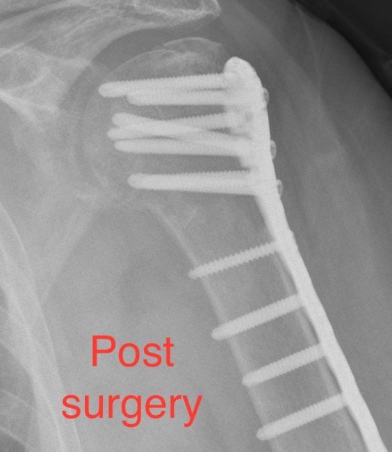
How do we treat these fractures? These fractures can be treated non-operatively in a sling if they are undisplaced (fracture ends not moved out from the original position) or minimally displaced. Once fracture shows signs of healing then shoulder physiotherapy may be started to gain good shoulder function. Serial plain radiographs may be required to make sure that fracture position is maintained. Surgical treatment may be required in displaced fractures however it also depends on patient’s general health and demands of daily life.
Shoulder arthritis and shoulder replacement
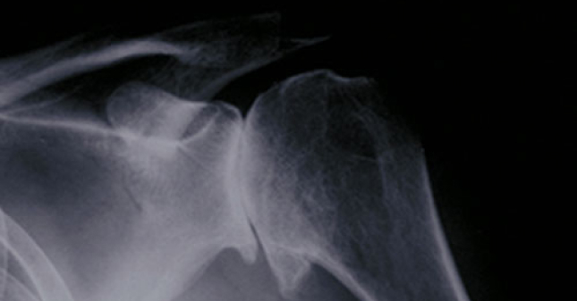
What is shoulder arthritis? Shoulder joint is a ball and socket type of joint and like other joints in the body they have smooth cartilage lining the joint articulating surfaces. In shoulder arthritis there is wear of this lining cartilage and this is similar to what you see in hip and knee joints although it is less common in shoulder joint. Loss of cartilage leads to bone rubbing against the bone and this leads to pain and loss of function.
Do I need more tests? Plain X-ray is performed followed by CT scan of the shoulder to understand the wear pattern of the joint more clearly.
What is the treatment? Painkillers are useful in managing shoulder arthritis initially. Cortisone injection into the shoulder joint may help to control symptoms for a short duration. Surgery involves a total shoulder replacement where the worn out socket is replaced with a plastic socket and the ball is replaced with a metal ball attached to a stem that goes into the arm bone (humerus). In some cases the socket is worn out too much that it may not be technically possible to replace it and then the ball part of the joint alone is replaced. This is called hemiarthroplasty or partial shoulder replacement. In some patients especially who are bit older, in addition to the arthritis the rotator cuff tendon may be torn or non functional. In such patients conventional shoulder replacement does not work and we offer reverse shoulder replacement (please see below).
Reverse shoulder replacement
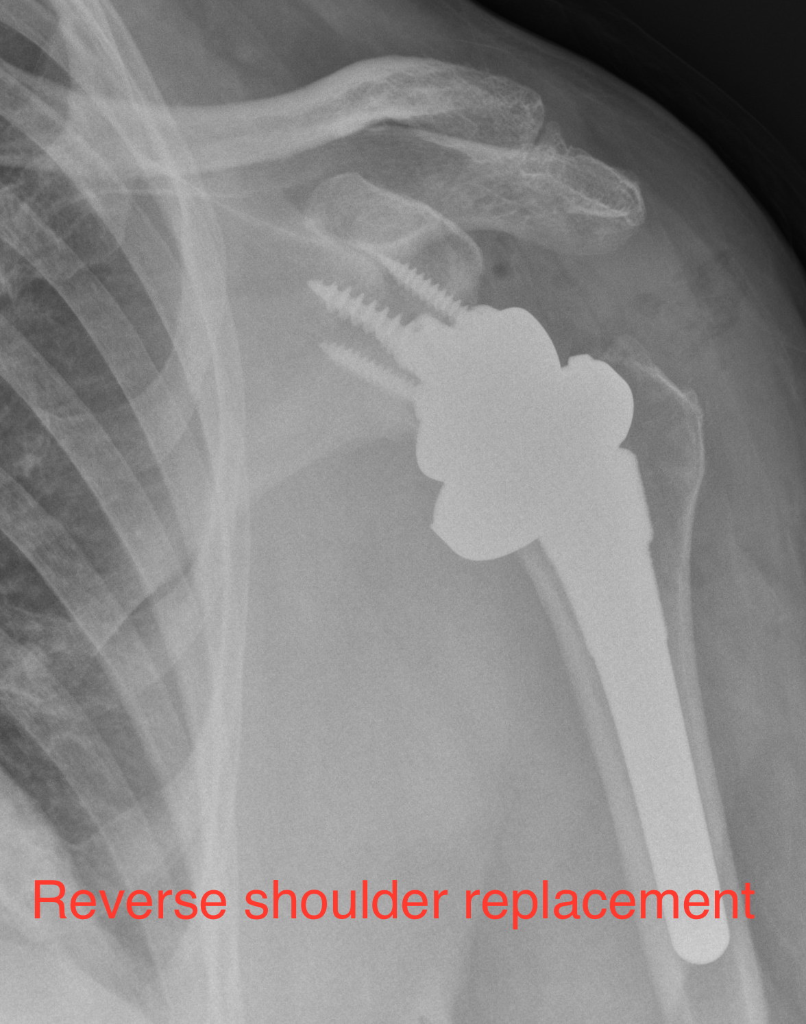
What is reverse shoulder replacement? In conventional shoulder replacement the ball of the shoulder joint is replaced by metal ball and the socket is replaced by plastic socket. This is reversed in a reverse shoulder replacement. A metal hemispherical ball replaces the socket and a plastic socket attached to the arm bone replaces the ball of the shoulder. This type of joint replacement utilises patient’s deltoid muscle for shoulder movement unlike conventional shoulder replacement, which relies on intact and functional rotator cuff muscle that envelopes the shoulder joint.
Who would benefit from reverse shoulder replacement? The primary indication for doing a reverse shoulder is in patients who have painful arthritis of the shoulder joint with completely torn rotator cuff (cuff tear arthropathy). This procedure is also done in older patients with irreparable rotator cuff tear who are in lot of pain and has severe weakness of the arm.
